
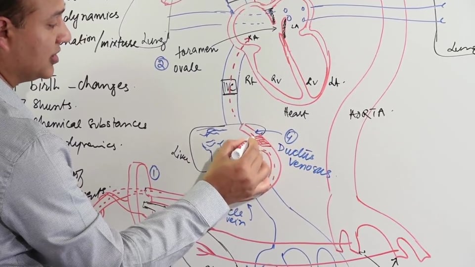
Development of the inferior venca cava. We will look at the sinus venosus and cardinal veins with the regression of some of the systems that eventually lead to the formation of the IVC.
STUDY NOTES:
DEVELOPMENT OF INFERIOR VENA CAVA
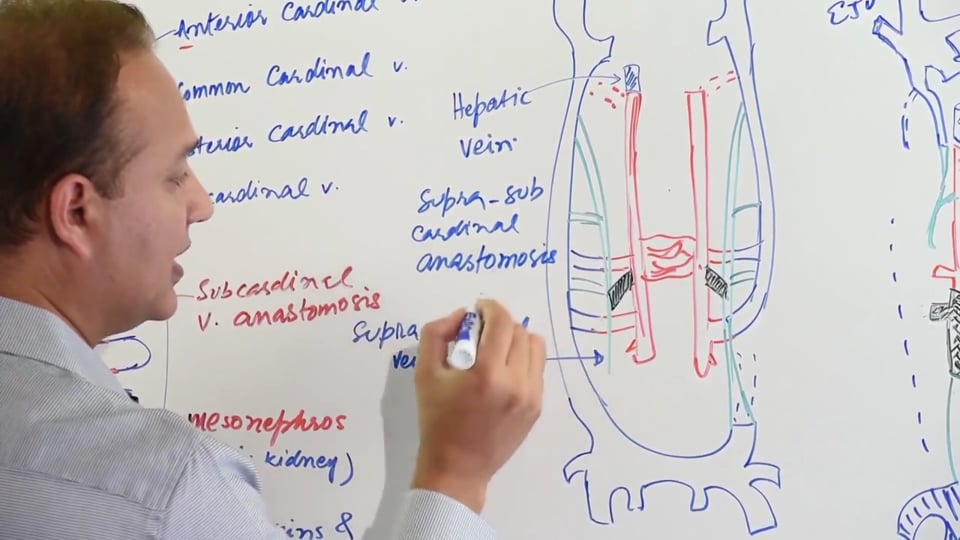
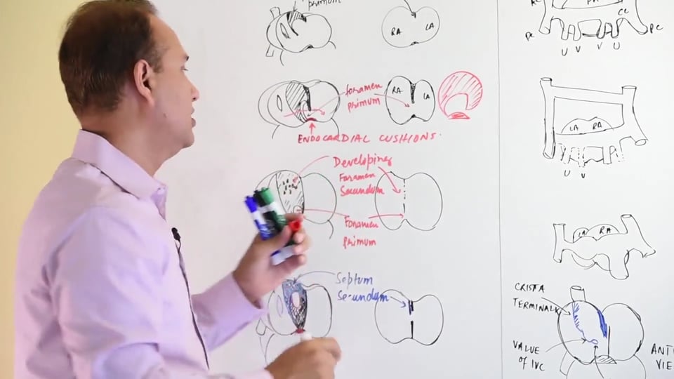
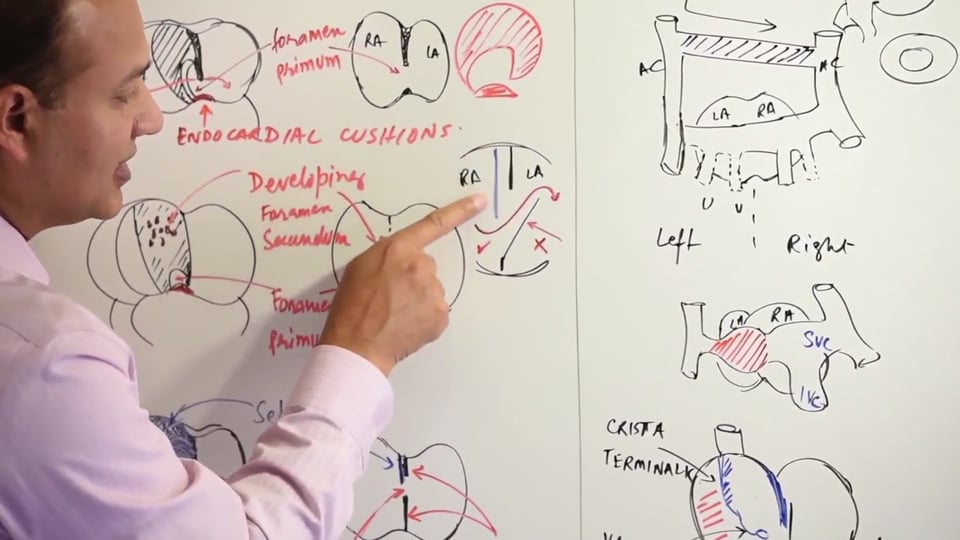
Sinus venosus forms the inflow tract of the primitive heart tube. On each side of the sinus venosus, the common cardinal veins open into it. Anterior and posterior cardinal veins combine to form the common cardinal vein on each side of the sinus venosus. The common cardinal vein on the right side forms part of the superior vena cava. Besides the common cardinal veins, the umbilical and the vitelline veins also drain into the sinus venosus. On the left side, these veins undergo specific remodeling. This results in formation of specific anastomosis on the left side, giving rise to left to right shunts thereby causing most of the blood to be received by the right side of the sinus venosus. Consequently, the veins of the right side start maturing and increase in size relative to their left side counterparts. As the sinus venosus also grows and matures, it becomes incorporated into the right side of the primitive atrium. Eventually the vitelline, common cardinal and the umbilical veins of the left side degenerate. The blood received by the left side of sinus venosus is greatly reduced and as a result the left side of the sinus venosus shrinks. Inferiorly the posterior cardinal veins anastomose together and form the iliac veins.
The formation of the inferior vena cava is contributed by three set of veins:
The anterior cardinal and the common cardinal veins on the left side give rise to brachiocephalic and the left subclavian vein which drain into the SVC. It is important to remember that, the umbilical vein on both sides and the vitelline vein on the left side, all tend to degenerate. The vitelline vein on the right side along with the common cardinal vein of the right side together form parts of the SVC.
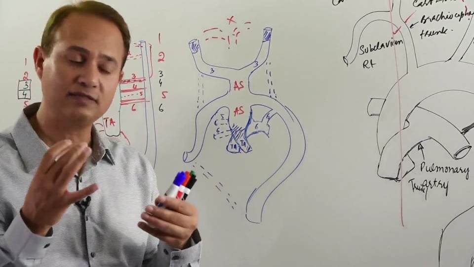
The subcardinal veins have an anastomosis in the middle which is referred to as subcardinal venous anastomosis. These subcardinal veins are also connected to the posterior cardinal vein and form another anastomosis which is referred to as the mesonephric shunt. Later on the inferior part of posterior cardinal vein on the left side starts degenerating, however, it's still connected to the subcardinal vein via the mesonephric shunt. Also, there are tiny buds arising from the subcardinal veins, these form parts of the future ovarian and spermatic veins.
Subcardinal veins on the right side separate from the posterior cardinal veins and are joined in by the hepatic veins. This will later form the initial parts of the IVC. At this point it's important to understand that an anastomosis forms between the supracardinal and the subcardinal veins, which is referred to as the supra-subcardinal anastomosis. This anastomosis becomes part of the IVC and later gives rise to the renal vein and parts of the spermatic veins. Over time the posterior cardinal vein on the right side also degenerates.
As the IVC is formed, it is composed of the following parts:
In this video we will learn about :
1. Venous flow of a fetus.
2. Sinus venosus contribution to IVC formation.
3. Cardinal veins formation to IVC formation.
4. Parts of IVC formed.
Following answers are created by ChatGPT. Occasionally the answer may be harmful, incorrect, false, misleading, incomplete, or limited in knowledge of world. Please contact your doctor for all healthcare decisions. Also, double check the answer provided by the AI below.
In addition to the presenter, following authors may have helped with the content writing, review, or approval:
ACCME Accreditation Statement
The DrBeen Corp is accredited by the Accreditation Council for Continuing Medical Education (ACCME) to
provide continuing medical education for physicians.
AMA Credit Designation Statement
The DrBeen Corp designates this enduring material for a maximum of 0.25 AMA PRA Category 1
Credits™.
Physicians should claim only the credit commensurate with the extent of their participation in the
activity.
In accordance with the disclosure policies of DrBeen Corp and the ACCME (Accreditation Council for
Continuing Medical Education), we are committed to upholding principles of balance, independence,
objectivity, and scientific rigor in all of our Continuing Medical Education (CME) and Continuing
Education (CE) activities. These policies include the careful management and mitigation of any relevant
financial relationships with organizations that are not eligible.
All members of the Activity Planning Committee and presenters have disclosed their relevant financial
relationships. The DrBeen Corp CE Committee has thoroughly reviewed these disclosures and determined
that these relationships are not deemed inappropriate in the context of their respective presentations.
Additionally, they are found to be consistent with the educational objectives and the integrity of the
activity.
| Faculty | Disclosures |
|---|---|
| Dr. Mobeen Syed | Author declares no conflict of interest. |
No credit card information needed.
 0.50 CME
Luis A Verduzco M.D.
 0.75 CME
Luis A Verduzco M.D.
Ahmed Zaafran, MD
 1.25 CME
Luis A Verduzco M.D.
Ahmed Zaafran, MD
 0.50 CME
Tatyana Travkina, MD
Ahmed Zaafran, MD
Ahmed Zaafran, MD
Ahmed Zaafran, MD
 0.75 CME
Tatyana Travkina, MD
Ahmed Zaafran, MD
Tatyana Travkina, MD
Ana Crawford M.D., M.Sc.
Ahmed Zaafran, MD
Ahmed Zaafran, MD
Ahmed Zaafran, MD
 1.25 CME
Dr. Mobeen Syed
Ahmed Zaafran, MD
 0.12 CME
Dr. Mobeen Syed

Ahmed Zaafran, MD
 1.25 CME
Dr. Mobeen Syed
Dr. Mobeen Syed
Dr. Mobeen Syed
 0.50 CME
Dr. Mobeen Syed
 0.75 CME
Dr. Mobeen Syed
Ahmed Zaafran, MD
 0.25 CME
Dr. Mobeen Syed
 0.16 CME
Dr. Mobeen Syed
 0.50 CME
Dr. Mobeen Syed
 0.16 CME
Dr. Mobeen Syed
Dr. Mobeen Syed
Dr. Mobeen Syed
 0.75 CME
Dr. Mobeen Syed
 0.50 CME
Dr. Mobeen Syed
 0.20 CME
Dr. Mobeen Syed
 0.20 CME
Dr. Mobeen Syed
 0.12 CME
Dr. Mobeen Syed
 0.09 CME
Dr. Mobeen Syed
 0.24 CME
Dr. Mobeen Syed
 0.25 CME
Dr. Mobeen Syed
 0.19 CME
Dr. Mobeen Syed
 0.08 CME
Dr. Mobeen Syed
 0.11 CME
Dr. Mobeen Syed
 0.09 CME
Dr. Mobeen Syed
 0.50 CME
Dr. Mobeen Syed
 1.00 CME
Dr. Mobeen Syed
 0.25 CME
Dr. Mobeen Syed
 0.50 CME
Dr. Mobeen Syed
 0.50 CME
Dr. Mobeen Syed
 0.50 CME
Dr. Mobeen Syed
 0.16 CME
Dr. Faraaz Bhatti
 0.50 CME
Dr. Mobeen Syed
 0.50 CME
Dr. Mobeen Syed
 0.50 CME
Dr. Mobeen Syed
 0.50 CME
Dr. Mobeen Syed

Dr. Mobeen Syed
 0.17 CME
Dr. Mobeen Syed
 0.50 CME
Dr. Mobeen Syed
Dr. Mobeen Syed
Dr. Mobeen Syed
Dr. Mobeen Syed
Dr. Mobeen Syed
Dr. Mobeen Syed
 0.50 CME
Dr. Mobeen Syed
Dr. Mobeen Syed
Dr. Mobeen Syed
Dr. Mobeen Syed
Dr. Mobeen Syed

Dr. Mobeen Syed

Dr. Mobeen Syed
 0.05 CME
Dr. Mobeen Syed
Dr. Mobeen Syed
 1.25 CME
Dr. Mobeen Syed
Dr. Mobeen Syed
Dr. Mobeen Syed
 0.50 CME
Dr. Mobeen Syed
 0.22 CME
Dr. Mobeen Syed
 0.50 CME
Dr. Mobeen Syed
 0.25 CME
Dr. Mobeen Syed
 0.13 CME
Dr. Mobeen Syed
 0.16 CME
Dr. Mobeen Syed
 0.16 CME
Dr. Mobeen Syed
 0.15 CME
Dr. Mobeen Syed
 0.15 CME
Dr. Mobeen Syed
 0.19 CME
Dr. Mobeen Syed
 0.75 CME
Dr. Mobeen Syed
 0.12 CME
Dr. Mobeen Syed
 0.16 CME
Dr. Mobeen Syed
 0.25 CME
Dr. Mobeen Syed
 0.24 CME
Dr. Mobeen Syed
 0.25 CME
Dr. Mobeen Syed
 0.19 CME
Dr. Mobeen Syed
 0.18 CME
Dr. Mobeen Syed
 0.50 CME
Dr. Mobeen Syed
 0.75 CME
Dr. Mobeen Syed
 0.50 CME
Dr. Mobeen Syed
 0.25 CME
Dr. Mobeen Syed
 0.50 CME
Dr. Mobeen Syed
 0.50 CME
Dr. Mobeen Syed
 0.25 CME
Dr. Mobeen Syed
 0.75 CME
Dr. Mobeen Syed
 0.13 CME
Dr. Mobeen Syed
 0.50 CME
Dr. Mobeen Syed
 0.25 CME
Dr. Mobeen Syed
 0.50 CME
Dr. Mobeen Syed
 0.50 CME
Dr. Mobeen Syed
 0.50 CME
Dr. Mobeen Syed
 0.25 CME
Dr. Mobeen Syed
 1.00 CME
Dr. Mobeen Syed
 0.25 CME
Dr. Mobeen Syed
 0.20 CME
Dr. Mobeen Syed
 0.25 CME
Dr. Mobeen Syed
 0.17 CME
Dr. Mobeen Syed
 0.25 CME
Dr. Mobeen Syed
 0.50 CME
Dr. Mobeen Syed
 0.25 CME
Dr. Mobeen Syed
0.19 CME
Dr. Mobeen Syed
 0.50 CME
Dr. Mobeen Syed
All information contained in and produced by DrBeen corp is provided for educational purposes only. This information should not be used for the diagnosis or treatment of any health problem or disease.
THIS INFORMATION IS NOT INTENDED TO REPLACE CLINICAL JUDGMENT OR GUIDE INDIVIDUAL PATIENT CARE IN ANY MANNER.
Click here for notice and disclaimer.
Write A New Comment
0 Comments